
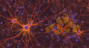
It has been known for some time that Alzheimer’s disease affects different regions of the brain in different ways and that tau, a protein known to malfunction, plays an important role in this disease. Normally, tau helps stabilize neurons, but in Alzheimer’s, it begins to fold incorrectly and becomes entangled in neurons. It spreads throughout the brain, forming toxic clumps that impair neuron function and eventually lead to cell death.
Researchers are Creating a Kind of “Google Maps” for the Movement of Tau Proteins in the Brains of Alzheimer’s Patients
Brain areas such as the entorhinal cortex and hippocampus are affected early by tau clumps, while other areas, such as the primary sensory cortex areas, remain resistant to the disease. To better understand this selective vulnerability (SV) or resistance (SR) to Alzheimer’s disease, researchers have conducted gene association and transgenic studies to identify Alzheimer’s risk genes. However, previous studies have not shown a clear link between the location of genetic risk factors and associated tau pathology.
A new study by researchers at UC San Francisco has now taken a major step toward answering this question by combining brain imaging, genetics, and advanced mathematical modeling into a powerful new approach. Published in Brain, the study reveals several distinct pathways through which risk genes influence susceptibility or resilience to Alzheimer’s disease. The study introduced a model for the spread of the disease called the “extended Network Diffusion Model” (eNDM). The researchers applied this model to brain scans of 196 people in various stages of Alzheimer’s disease. They subtracted the model’s predictions from the scan results. The remaining values, called “residual tau,” pointed to areas where factors other than brain connections influence tau formation—in this case, genes.
Using gene expression maps of the brain from the Allen Human Brain Atlas, the researchers tested the extent to which Alzheimer’s risk genes explain the patterns of both actual and residual tau. This allowed them to separate genetic effects that act with or independently of the connections in the brain. “We think of our model as Google Maps for tau,” said the study’s senior author, Ashish Raj, PhD, professor of radiology and biomedical imaging at UCSF. “It uses real data about the connections in the brains of healthy people to predict where the protein is likely to migrate next.”
Genes that Make Certain Parts of the Brain More or Less Susceptible to the Disease
The research team discovered four different types of genes, depending on the extent to which and how they predict tau: Network-aligned vulnerability (SV-NA), which are genes that promote the spread of tau along connections in the brain; Network-independent vulnerability (SV-NI), which are genes that promote tau deposition in a way that is not related to connectivity; Network-aligned resilience (SR-NA), i.e., genes that protect regions that are otherwise tau hotspots; and Network-independent resilience (SR-NI), i.e., genes that provide protection outside the usual network pathway—like hidden shields in unlikely places.
“Genes associated with vulnerability deal with stress, metabolism, and cell death; genes associated with resilience are involved in the immune response and the clearance of amyloid beta, another trigger of Alzheimer’s,” explained the study’s first author, Dr. Chaitali Anand, a postdoctoral fellow at UCSF. “Essentially, the genes that make certain parts of the brain more or less susceptible to Alzheimer’s perform different tasks—some control the movement of tau, others are responsible for internal defense mechanisms or cleaning systems.”
This research builds on another recent UCSF study in mice published in Alzheimer’s & Dementia, which showed that tau does not migrate randomly or diffuse passively, but follows the brain’s wiring pathways with a distinct directional preference. Using a system of differential equations called the Network Diffusion Model (NDM), the research team was able to demonstrate the dynamics of tau propagation between connected brain regions, refuting the traditional view that tau simply spreads through diffusion in the extracellular space or by leaking out of dying neurons.
Helpful in Identifying Potential Targets for Intervention
“Our research has shown that tau propagates transsynaptically by migrating along axonal projections, driven by active transport processes rather than passive diffusion, and utilizing active neural pathways in the preferred retrograde direction,” said Justin Torok, PhD, postdoctoral fellow in the Raj lab. In the current study, network-based analyses complemented existing approaches to validate and identify gene-based determinants of selective susceptibility and resilience. Genes that respond independently of the network have different biological functions than genes that respond in concert with the network.
This study offers a promising way forward: one that connects biology and brain maps to a smarter strategy for understanding and ultimately curing Alzheimer’s disease. The findings provide new insights into signs of susceptibility to Alzheimer’s and may prove helpful in identifying potential targets for intervention.
Related Posts
-

Alzheimer's disease, the most common form of dementia in the elderly, impairs cognitive ability. That…
-
Researchers at the Medical College of Georgia at Augusta University want to better understand why…
-
Memory loss, confusion, language problems – Alzheimer's disease is the most common cause of dementia…