

Japanese researchers have discovered a sulfur-containing compound that could significantly enhance the effectiveness of an important endogenous muscle repair protein. In laboratory experiments, the substance—called lipoic acid trisulfide (LASSS)—improved the protein’s binding ability and could thus influence a key mechanism of muscle regeneration. The findings open up a new research approach in the fight against age-related muscle wasting. Further studies are needed to determine whether this discovery can lead to future treatment options for humans.
When Natural Muscle Repair Declines
As we age, muscles don’t just lose strength; their ability to repair damage also declines. Japanese scientists have now identified a potential approach to strengthen a key natural repair mechanism in the body. A specific sulfur-containing compound could play a surprising role in this process.
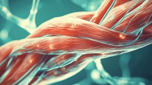
Muscles are under constant strain throughout a person’s life. They enable movement, stabilize the body, and constantly respond to stress. At the same time, they have the ability to regenerate damaged tissue on their own. However, as we age, this repair mechanism no longer functions as reliably as it does in youth. The consequences can be far-reaching: muscle tissue loses its performance capacity, recovery from injuries takes longer, and physical endurance declines. The risk of age-related muscle loss also increases.
A research team at Kyushu University in Japan has investigated a biological mechanism that could be crucial to this process. The focus is on a protein called Hepatocyte Growth Factor (HGF). It plays an important role in activating the cells responsible for repairing and regenerating muscle tissue. The scientists discovered that the function of this protein can potentially be significantly improved using a sulfur-containing compound.
An Important Repair Protein Loses Its Effectiveness With Age
So-called satellite cells are necessary for muscles to regenerate damaged tissue. These specialized stem cells are located in the immediate vicinity of muscle fibers and play a central role in muscle regeneration. When activated by injury or physical stress, they begin to multiply and support the repair of damaged muscle fibers. The protein Hepatocyte Growth Factor (HGF) plays a crucial role in this process. It binds to a receptor called c-Met on the surface of the satellite cells, thereby triggering signals that set the regeneration process in motion. Without this signal transmission, the stem cells cannot fully perform their repair function.
However, HGF can lose its effectiveness due to chemical modifications. One such modification is a process known as nitration. In this process, a chemical group is attached to specific sites on the protein, which can alter its structure and function. The Japanese researchers had already determined that two specific sites on the HGF protein are particularly susceptible to this process. If these sites are altered, the protein has difficulty binding to the c-Met receptor. This may impair the activation of satellite cells. In other words, while the body can still produce this important repair protein, its biological efficacy may be limited. The researchers suspect that such changes could be a previously overlooked factor in declining muscle regeneration.
Earlier Studies Had Already Provided Important Clues
Scientists had already determined in earlier studies that the chemical alteration of the HGF protein could indeed be linked to the aging process. In a study published in 2023 in the journal *Aging Cell*, the research team examined muscle tissue from rats of different ages. They found that the nitration of the HGF protein increased with age. Certain fast-twitch muscle fibers were particularly affected.
Further laboratory tests confirmed that this chemical change can significantly impair the protein’s binding to its receptor, c-Met. This could disrupt the activation of muscle stem cells, which in turn hinders the natural regeneration of muscle tissue. Another study from 2024 therefore explored ways to prevent this harmful change. The current study using LASSS builds on these research findings and, for the first time, takes the approach of not only protecting the HGF protein but actually increasing its binding capacity beyond its original level
Sulfur Compound Alters the Protein’s Properties
To determine how this loss of function can be prevented, the team investigated two sulfur-containing compounds with antioxidant properties: glutathione trisulfide (GSSSG) and lipoic acid trisulfide (LASSS). Both substances belong to the group of trisulfides. A characteristic feature of these compounds is a bond consisting of three sulfur atoms. They can participate in so-called redox reactions and thereby influence certain chemical processes in cells.
In laboratory experiments, both compounds were able to reduce the undesirable nitration of the HGF protein. However, it initially became apparent that protection against this chemical modification alone was not sufficient to fully restore the protein’s original binding capacity. Only at a higher concentration of LASSS did the scientists observe an effect they apparently had not anticipated: The protein was suddenly able to bind significantly better to its receptor, c-Met.
The binding capacity was more than twice as high compared to untreated HGF. At the same time, there was increased resistance to the loss of function that can result from nitration. Particularly interesting: This additional effect occurred with LASSS, but not with the compound GSSSG, which was also studied.
Researchers Refer to a Kind of “Super HGF”
The scientists suspect that LASSS not only acts as an antioxidant but also interacts directly with the HGF protein. In doing so, the sulfur compound could alter its three-dimensional structure and thus improve its ability to bind to the c-Met receptor. A protein’s three-dimensional structure is crucial to its function. Even minor changes can influence how well it interacts with other molecules. In the case of HGF, this could result in the protein transmitting its signals to muscle stem cells more effectively. The researchers therefore refer to it as a kind of “super HGF.” In laboratory experiments, the protein treated with LASSS exhibited more than twice the binding affinity for the c-Met receptor compared to the untreated version. However, whether this increased activity actually leads to improved muscle regeneration still needs to be clarified in further studies.
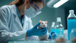
Following the laboratory experiments, the researchers wanted to know whether LASSS could also exert a protective effect within living muscle tissue. To do this, they used mice in which normal muscle load had been reduced via a procedure known as tail suspension. This experimental method is used to investigate changes that can occur during prolonged physical inactivity, including the breakdown of muscle tissue. The animals that had received LASSS prior to the experiment showed significantly lower nitration of the HGF protein than the untreated control group.
The researchers thus found initial evidence that the compound can influence the chemical changes in the repair protein not only under controlled laboratory conditions but also within a living organism. However, an important limitation must be noted: The treatment did not demonstrably result in the mice developing more muscle mass or greater muscle strength. The study primarily investigated whether LASSS can protect the HGF protein from certain chemical changes. The second sulfur compound, GSSSG, also did not exhibit the same protective effect in this experiment.
Could this Discovery Help Counteract Muscle Wasting in the Future?
In the long term, this discovery could open up new possibilities for addressing age-related muscle wasting. This is because, as we age, the body loses not only muscle mass and strength but also some of its natural ability to repair damaged muscle tissue. Similar problems can arise after prolonged periods of physical inactivity, such as after surgery, during prolonged bed rest, or during severe illness. In such situations, muscle loss can be further accelerated.
The Japanese researchers’ approach differs from previous strategies, which have primarily focused on building new muscle mass. Instead, the goal is to support the body’s existing repair mechanism. The focus is on the HGF protein, whose function can be impaired by chemical changes. If LASSS can successfully prevent these changes while simultaneously improving the protein’s binding capacity, this could support the muscles’ natural regeneration processes. Should further studies confirm the current findings, LASSS could potentially serve as an interesting starting point for developing new therapeutic approaches to combat age-related muscle wasting. In the long term, applications could also be conceivable for people whose muscles have been weakened by prolonged immobility or disease.
The researchers also see potential applications in veterinary medicine. Older dogs and cats, in particular, could benefit in the future from new approaches aimed at limiting age-related loss of muscle tissue. Until then, however, further studies are necessary. Above all, it must be clarified whether LASSS actually improves muscle regeneration, maintains muscle strength, and can be used safely over the long term. The results to date provide an interesting approach toward this end, but do not yet demonstrate a therapeutic effect in humans
There is Still a Long Way to Go Before Human Application
As promising as the results may seem, numerous questions remain unanswered before a potential medical application can be considered. In particular, it must be determined whether LASSS can actually improve muscle regeneration in older organisms. This requires further studies in aging animals. Potential side effects, safety, and appropriate dosing must also be investigated.
Furthermore, it has not yet been proven that the enhanced binding of the HGF protein actually leads to increased muscle strength, greater muscle mass, or improved physical performance in humans. The study therefore does not yet provide a basis for a new treatment for muscle wasting. However, it does highlight an interesting biological approach: It may be possible to improve age-related declines in muscle repair by protecting a key endogenous signaling protein from harmful changes. Precisely because the approach builds on an existing repair mechanism, it could prove valuable for future research on aging and regeneration.