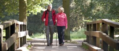
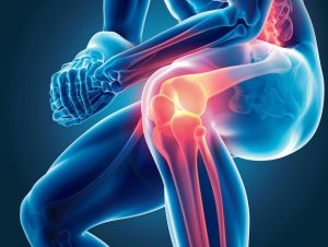
Almost a quarter of all people over the age of 40 suffer from painful osteoarthritis, making it one of the most common causes of disability in adults. Osteoarthritis leads to wear and tear of the cartilage that cushions the joints, and there is currently no way to reverse this damage: the only option is to treat the pain with medication and, ultimately, joint replacement. Researchers at the University of Utah, New York University, and Stanford University are now demonstrating the potential of another option: changing the way people walk. By making a small adjustment to the angle of their feet when walking, participants in a one-year randomized controlled trial achieved pain relief equivalent to that achieved with medication.
Crucially, these participants also experienced less knee cartilage loss during this period compared to a group that received a placebo treatment. These results were published in The Lancet Rheumatology and co-led by Scott Uhlrich of the John and Marcia Price College of Engineering in Utah. They come from the first placebo-controlled study to demonstrate the effectiveness of a biomechanical intervention in osteoarthritis.
How Gait can Influence Osteoarthritis
“We know that in people with osteoarthritis, increased stress on the knee accelerates the progression of the disease and that changing the angle of the foot can reduce knee stress,” said Uhlrich, assistant professor of mechanical engineering. “So the idea of a biomechanical intervention is not new, but there have been no randomized, placebo-controlled studies to prove its effectiveness.” With support from the National Institutes of Health and other federal agencies, the researchers specifically studied patients with mild to moderate osteoarthritis in the medial compartment of the knee—the inner side of the leg—which tends to bear more weight than the lateral, outer compartment.
This form of osteoarthritis is the most common, but the ideal foot angle for relieving the medial side of the knee varies from person to person, depending on their natural gait and how it changes when a new gait is adopted. “In previous studies, the same measure was prescribed to all individuals, which meant that in some cases the joint load was not reduced or even increased,” said Uhlrich. “We took a personalized approach to develop a new gait pattern for each person, which improved knee relief and likely contributed to the positive effects on pain and cartilage that we observed.”
During their first two visits, participants underwent a baseline MRI scan and practiced walking on a pressure-sensitive treadmill while motion capture cameras recorded the mechanics of their gait. This allowed the researchers to determine whether turning the toes inward or outward reduced the load more and whether an adjustment of 5° or 10° was ideal. This personalized analysis also excluded potential participants who would not benefit from the intervention because none of the foot angle changes could reduce the load on their knees. These subjects had been included in previous studies, which may have contributed to the inconclusive pain results of those studies. In addition, after the initial intake sessions, half of the 68 participants were assigned to a sham treatment group to control for the placebo effect. These participants were prescribed foot angles that were actually identical to their natural gait. Conversely, the individuals in the intervention group were prescribed the foot angle change that maximally reduced their knee load.
Less Pain and Better Cartilage Health
Participants in both groups returned to the lab for six weekly training sessions, where they received biofeedback—vibrations from a device worn on the shin—that helped them maintain the prescribed foot angle while walking on the lab treadmill. After the six-week training phase, participants were encouraged to practice their new gait for at least 20 minutes a day until it became second nature. Regular follow-up examinations showed that participants maintained the prescribed foot angle to within one degree on average. After one year, all participants reported on their experiences with knee pain and underwent a second MRI scan to quantitatively assess the damage to their knee cartilage. “The reported pain reduction compared to the placebo group was between what you would expect from an over-the-counter medication such as ibuprofen and an anesthetic such as Oxycontin,” said Uhlrich. “The MRI scans also allowed us to see a slower deterioration of a marker for cartilage health in the intervention group, which was very exciting.”
Later Knee Surgery
Beyond the quantitative measures of efficacy, study participants were enthusiastic about both the approach and the results. One participant said, “I don’t have to take any medication or wear a device… it’s just part of my body now that will be with me for the rest of my life, and I’m very happy about that.” The ability of participants to adhere to the intervention over a long period of time is one of its potential benefits. “Especially for people in their thirties, forties, or fifties, osteoarthritis can mean decades of pain management before joint replacement is recommended.
This intervention could help close that huge treatment gap. Before this measure can be used clinically, the gait training process needs to be optimized. The motion capture technology used to create the original foot angle prescription is expensive and time-consuming. The researchers envision that this measure will eventually be used in a physical therapy practice, with training taking place during a walk in the neighborhood. Further studies on this approach are needed before the intervention can be made available to the general public.
Related Posts
-

There is new research to show that fasting triggers a molecule that can help delay…
-
Adults aren't eating enough fiber. Considering that fiber could play a key role in reducing…
-
As you get older, it is important to stay active and keep your body moving.…