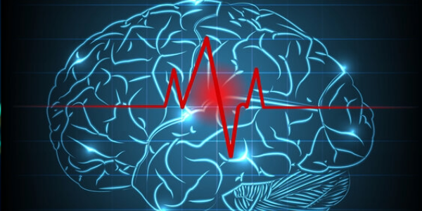
A new study in The Lancet Digital Health suggests that the brain may respond to a stroke in surprising ways. Researchers at the USC Mark and Mary Stevens Neuroimaging and Informatics Institute (Stevens INI) found that people with severe physical impairments following a stroke may show signs of a “younger” brain structure in undamaged areas. This appears to demonstrate how the brain adapts and reorganizes itself after damage.
What Happens During a Stroke?
A stroke occurs when the blood supply to a part of the brain is suddenly interrupted or severely restricted. As a result, the nerve cells no longer receive enough oxygen and nutrients, causing them to begin dying after just a few minutes. In most cases, a stroke is caused by a blocked blood vessel, such as a blood clot that obstructs blood flow. Less commonly, the cause is a blood vessel in the brain bursting and causing bleeding that damages the surrounding tissue.
The consequences depend on which region of the brain is affected. Often, sudden paralysis or weakness occurs on one side of the body, for example in the arm or face, which can manifest as a drooping corner of the mouth. Speech problems, visual disturbances, dizziness, or severe headaches may also occur. Because the brain is very sensitive and damaged nerve cells rarely regenerate, every minute counts in the event of a stroke. The faster medical treatment is provided, the greater the chance of preventing or reducing permanent damage. That is why a stroke is always a medical emergency requiring immediate assistance.
AI Reveals Brain Restructuring After a Stroke
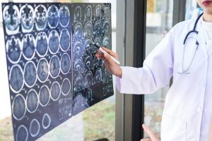
The research was conducted as part of the ENIGMA (Enhancing NeuroImaging Genetics through Meta-Analysis) working group on stroke recovery. The scientists analyzed brain scans from more than 500 stroke patients collected at 34 research centers in eight countries. Using deep learning models trained on tens of thousands of MRI scans, the team estimated the “brain age” of different regions in each hemisphere and examined how a stroke affects both structure and recovery. “We found that more severe strokes accelerate aging in the damaged hemisphere, yet paradoxically make the opposite side of the brain appear younger,” said Dr. Hosung Kim, associate professor of research neurology at the Keck School of Medicine of USC and one of the study’s lead authors. “This pattern suggests that the brain may be reorganizing itself and essentially rejuvenating the undamaged networks to compensate for the loss of function.”
To conduct the analysis, the researchers used a type of artificial intelligence known as a graph convolutional network. This system estimated the biological age of 18 brain regions based on MRI data. They then compared this predicted age with each person’s actual age—a measure known as “brain-predicted age difference” (brain-PAD), which serves as an indicator of brain health.
When these measurements of brain age were compared with motor function scores, a clear pattern emerged. Stroke patients with severe movement impairments exhibited a younger-than-expected brain age in regions opposite the injury site, even after more than six months of rehabilitation. This effect was particularly pronounced in the frontoparietal network, which plays a key role in movement planning, attention, and coordination.
“These results suggest that undamaged regions on the opposite side of the brain can adapt to compensate when stroke damage leads to a greater loss of movement,” explained Kim. “We observed this in the contralateral frontoparietal network, which exhibited a ‘more youthful’ pattern and is known to support movement planning, attention, and coordination. Rather than suggesting a full recovery of motor function, this pattern may reflect the brain’s attempt to adapt when the damaged motor system can no longer function normally. This offers us a new perspective on neuroplasticity that could not be captured using conventional imaging techniques.”
Toward Personalized Stroke Recovery
The study drew on ENIGMA, a global collaboration that brings together data from more than 50 countries to better understand the brain under various conditions. By standardizing MRI data and clinical information from many research groups, the team created the largest neuroimaging dataset of its kind on stroke.
“By combining data from hundreds of stroke patients worldwide and using state-of-the-art AI, we can identify subtle patterns of brain restructuring that remained invisible in smaller studies. These insights into regionally distinct brain aging processes in chronic stroke could ultimately serve as the basis for personalized rehabilitation strategies,” said Dr. Arthur W. Toga, director of the Stevens INI and Provost Professor at USC.
The researchers plan to continue this work by following patients over an extended period—from the early stages after a stroke through to long-term recovery. Tracking the development of aging patterns and structural changes in the brain could help clinicians tailor treatments to each person’s individual recovery process, with the goal of improving outcomes and quality of life. The study “Deep-learning prediction of MRI-based regional brain aging reveals contralateral neuroplasticity associated with severe motor impairments in chronic stroke.”
Related Posts
-

It has been known for some time that Alzheimer's disease affects different regions of the…
-
The accumulation of tau protein in the brain is a characteristic feature of Alzheimer's disease.…
-
Brain health is a significant concern for many people. Various factors can contribute to brain…