Many eye diseases do not lead to vision loss suddenly, but rather over the course of years. Progressive damage to the retina plays a central role in this process. Scientists are therefore increasingly trying to understand how this sensitive tissue reacts to such damage—and whether its own protective mechanisms can be specifically supported. A research team at the Scripps Research Institute, in collaboration with scientists from the University of California, San Diego, and the Lowy Medical Research Institute, has identified a molecule that could play an important role in this process: erucamide. The study, published in Nature Neuroscience, suggests that erucamide is involved in communication between different cell types in the retina. When erucamide levels drop during retinal degeneration, the tissue’s natural protective mechanism appears to be weakened at the same time.
When Photoreceptors Die
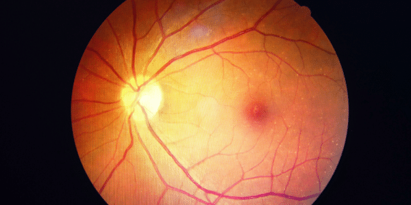
As we age, the eye also changes. The retina, which detects light stimuli inside the eye and converts them into electrical signals for the brain, is particularly vulnerable to stress. Certain age-related changes can contribute to the sensitive cells of the retina becoming more susceptible to damage. This is particularly relevant in conditions such as age-related macular degeneration (AMD), one of the most common causes of progressive loss of central vision in older adults. However, other retinal diseases can also lead to the gradual damage of photoreceptors and a decline in visual performance.
As we age, therefore, the focus shifts not only to why retinal cells are damaged, but also to how the eye responds to this damage. to keep them functioning. This current research focuses precisely on this natural protective mechanism. The results could provide new insights into how the eye’s natural defense mechanisms might be harnessed to slow the progressive damage to the retina.
The retina consists of a complex interplay of different cell types. In addition to the photoreceptors—which are crucial for vision—nerve cells, glial cells, blood vessels, and immune cells are also involved. These different components must work closely together to ensure that the retina is adequately supplied with oxygen and nutrients and can maintain its function. This interaction is often referred to as the neurovascular unit.
In diseases such as age-related macular degeneration, diabetic retinopathy, or retinitis pigmentosa, this balance can become increasingly disrupted. The photoreceptors are damaged and gradually die off. This also results in a loss of visual function. The researchers therefore sought to determine whether the retina releases specific chemical signals in response to such damage that help the remaining tissue protect itself.
In Search of a Protective Signal
Earlier studies by the research team provided an important clue. In those studies, transplanted retinal cells derived from stem cells appeared to have a protective effect on degenerating tissue. Remarkably, this effect persisted to some extent even after the transplanted cells themselves had already disappeared. The scientists therefore reasoned that the cells might release certain signaling molecules that trigger a longer-lasting response in the tissue.
To identify such molecules, the researchers examined the chemical composition of retinal tissue in various preclinical models. To do so, they used mass spectrometric metabolomics, a technique that allows for the simultaneous detection of numerous small molecules. Erucamide stood out in particular.
The levels of this molecule decreased significantly as the photoreceptors became increasingly damaged. This led the scientists to wonder whether the decline in erucamide was merely a side effect of the disease—or whether the molecule itself plays a role in the retina’s response.
What Happens When Erucamide is Reintroduced?
To investigate this question, the researchers specifically reintroduced erucamide into the retina. To do so, they used porous silicon nanoparticles that served as a delivery system. This was necessary because erucamide is hydrophobic and therefore dissolves poorly in water. The nanoparticles enabled a more stable and controlled distribution of the molecule within the eye.

Subsequent investigations provided important insight into the mechanism of action. Erucamid did not appear to directly protect the damaged photoreceptors. Instead, the molecule influenced certain immune cells in the retina, known as CD11b⁺ myeloid cells. These cells are involved in responses to injury and can send out signals that are important for tissue preservation.
TMEM19 as a Possible Component of the Signaling Pathway
The researchers also identified the protein TMEM19 as a binding partner of erucamide. When the level of TMEM19 was reduced, the response of the myeloid cells to erucamide also changed. At the same time, the observed protective effect of the molecule was lost.
The results thus suggest that erucamide acts via a signaling pathway involving TMEM19 and myeloid immune cells. Upon activation, the cells in turn release signals associated with the stabilization of the neurovascular environment. This could support both nerve cells and the blood vessels that supply them.
No Cure – But Possibly Greater Stability
It is important to note that Erucamid did not reverse retinal degeneration in the models studied. Rather, the results suggest that the molecule can slow certain degenerative processes and help preserve the structure and function of the remaining retinal tissue for longer.
It is precisely this approach that could be of interest to researchers. Instead of exclusively treating the already damaged photoreceptors directly, a future therapy could aim to make the environment surrounding the nerve cells more resilient to damage. This is because photoreceptors do not function in isolation: their function and survival depend on a well-coordinated interaction with blood vessels, glial cells, and immune cells. If this delicate balance is disrupted by a disease, damage to the retina can worsen.
Based on the results so far, erucamide could help stabilize this environment. The molecule appears to activate certain immune cells, which in turn send out signals that support the nerve cells and blood vessels of the retina. This could enable the remaining cells to better cope with the stresses of a progressive disease. The goal, therefore, would not be to restore photoreceptors that have already died, but rather to protect the remaining healthy tissue for as long as possible and maintain its function.
Still a Long Way to Go Before a Treatment
Whether erucamide can one day actually be used as a medication for retinal diseases remains to be seen. One of the challenges lies in administering the molecule. Due to its poor water solubility, it cannot be easily used with conventional, water-based formulations for the eye.
The scientists therefore plan to investigate, among other things, modified variants of erucamide. These could potentially have a stronger or longer-lasting effect. In addition, other related lipid molecules will be investigated. Above all, the study provides an interesting insight into how the retina attempts to defend itself against progressive damage.
Erucamid could be part of an existing biological defense system. Instead of introducing a completely new mechanism into the eye, a future treatment could potentially amplify a signaling pathway that the retina itself uses. However, further research is needed before this can lead to a concrete therapy. Nevertheless, the research shows that the body’s own responses to retinal damage may have greater therapeutic potential than previously thought. Erucamide could thus provide another starting point for better understanding how retinal degeneration develops—and how the loss of functional tissue might be slowed.
Related Posts
-

As men and women age, joint damage is a common issue that can occur in…
-
Although the liver is one of the body's most resilient organs, it is still susceptible…
-
According to the World Health Organization, almost 530 million people worldwide were living with osteoarthritis…