
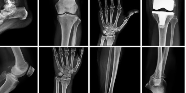
Bone and skeletal injuries are one of the leading causes of long-term disability worldwide. Researchers at Lund University in Sweden have developed a cell-free cartilage structure designed to help the body repair bone damage. According to the study, this artificially produced graft can promote bone healing without triggering strong immune responses. The method has already been successfully tested in animal models, and the researchers are preparing to evaluate the approach in human trials.
Major Bone Injuries Often Require Transplants
When large portions of bone are destroyed or removed, the body may have difficulty repairing the damage on its own. This can occur following cancer treatment, in cases of severe joint diseases such as rheumatoid arthritis and osteoarthritis, or in cases of severe infections. In these cases, a bone transplant is often necessary to restore structure and function.
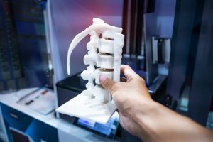
Researchers estimate that more than two million people worldwide require bone transplants each year. Current treatment methods typically rely on the use of the patient’s own tissue or cells to rebuild the bone. While this approach can be effective, it is costly, time-consuming, and can place an additional physical burden on patients. Furthermore, as the researchers note, it contributes to rising healthcare costs.
Toward a Universal Technology for Bone Regeneration
“Patient-specific grafts are both costly and time-consuming and do not always lead to success. A universal approach to tissue engineering with a reproducible manufacturing process offers significant advantages. In our study, we present exactly such a method and highlight important progress toward a non-patient-specific technology,” said Alejandro Garcia Garcia, a research associate in the field of molecular skeletal biology at Lund University. To develop this new method, the team first grew cartilage tissue in the lab. They then removed all living cells from it in a process known as decellularization. This step leaves behind the extracellular matrix—the natural scaffold that surrounds the cells in the tissue and provides both structural support and biological signals.
Since this scaffold remains intact, it continues to contain growth factors that can guide the body’s own cells. When the remaining cartilage structure is placed at a site of injury, it can act as a blueprint that helps the body rebuild the damaged bone step by step.
“The cartilage structure we have developed is based on stable, well-controlled, and reproducible cell lines and can stimulate bone formation without triggering strong immune reactions. We demonstrate that it is possible to produce a ready-made, so-called ‘off-the-shelf’ graft that interacts with the immune system and can repair large bone defects. Since the material can be manufactured and stored in advance, we see this as an important step toward the future clinical use of human bone tissue grafts,” said Paul Bourgine, who led the study. He is an associate professor and researcher in the field of molecular skeletal biology at Lund University.
Preparing for Clinical Trials in Humans
A key advantage of this technology is that the cartilage scaffold can be manufactured in advance and used in many patients without the need for individual customization. In the next phase of research, the focus will be on evaluating the method in humans, while simultaneously scaling up and standardizing production.
“The next step is to decide which types of injuries we want to test this on first, such as severe defects in the long bones of the arms and legs. At the same time, we need to prepare the documentation required for ethical review and regulatory approval to conduct clinical trials. At the same time, we are establishing a manufacturing process that can be carried out on a larger scale while consistently ensuring the same high level of quality and safety,” said Alejandro Garcia Garcia.
One Injection Reversed Osteoarthritis Within a Few Weeks
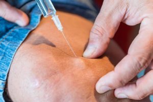
Researchers at the University of Colorado Boulder, CU Anschutz, and Colorado State University have developed a series of experimental treatments that could help aging and damaged joints regenerate on their own within a few weeks. The therapies have shown promising results in animal studies: they were able to reverse signs of osteoarthritis and restore the health of the joints. The new approaches include a regenerative injection administered directly into a joint, as well as a biomaterial-based repair system that stimulates the body’s own cells to rebuild damaged cartilage.
“Within two years, we’ve gone from a bold idea to developing these therapies and proving that they can reverse osteoarthritis in animals,” said lead researcher Stephanie Bryant, a professor of chemical and bioengineering at CU Boulder. “Our goal is not only to treat pain and halt the progression of the disease, but to defeat this disease once and for all.”
A New Approach to Treating Osteoarthritis
Osteoarthritis affects about one in six people worldwide over the age of 30. The disease leads to a gradual breakdown of cartilage, the cushioning tissue that prevents bones from rubbing against each other. As the disease progresses, it can also damage the bone, alter the joint structure, and make everyday movements increasingly painful. Current treatment options are limited. Most patients either manage their symptoms through pain management or eventually undergo joint replacement surgery. There is currently no cure for the disease. Researchers in Colorado are pursuing two different strategies to change this situation.
One treatment method uses a drug already approved by the Food and Drug Administration in a new application. Bryant and her colleagues developed a patented particle-delivery system that can be injected into a joint, where it releases regular doses of the drug over several months. For patients with more extensive damage to cartilage or bone, the team developed a separate therapy consisting of genetically engineered proteins. The material is administered arthroscopically, hardens at the injection site, and attracts the body’s own progenitor cells to repair the damaged area.
Rapid Joint Repair in Animal Studies
When the researchers tested the injectable treatment on animals with osteoarthritis and joint injuries, the affected joints returned to a healthy state within four to eight weeks. The repair material also delivered impressive results. According to Bryant, filling defects in cartilage or bone led to “complete regeneration and repair of the defect.” The therapies also demonstrated regenerative effects in human cells derived from patients who had undergone joint replacement surgery. NITRO was the first program launched by ARPA-H and was established to “develop minimally invasive therapeutics that fully regenerate damaged joints.” Two years ago, the program awarded the Colorado-based team up to $33.5 million—subject to positive results—to pursue this goal. With the successful completion of the first phase, the researchers have now advanced to the second phase.
Dr. Evalina Burger, professor and chair of the Department of Orthopedics at CU Anschutz, explained that osteoarthritis affects people from all walks of life. She has seen how the disease impacts grandparents who struggle with simple daily tasks due to shoulder pain, as well as athletes who are forced to give up running, ice hockey, and other activities because of knee or back problems. “Currently, many patients have to choose between major, expensive surgery or nothing at all. There isn’t much in between,” said Burger, who has been following the team’s research with interest. “That’s why ARPA-H is so important.”
Burger and Bryant envision a future in which people with early-stage osteoarthritis could receive an affordable, one-time treatment that keeps their joints healthy for years to come. For patients with localized cartilage or bone injuries, the damaged tissue could potentially be repaired in a single doctor’s visit, allowing them to recover quickly. The researchers plan to publish the results of their animal study in a peer-reviewed journal later this year. They have also founded the company Renovare Therapeutics Inc. to bring the technology to market. If future research continues to yield positive results, clinical trials could begin as early as 18 months from now, according to Bryant.